Pediatrics NUR 335 Exam Questions

Access The Exact Questions for Pediatrics NUR 335 Exam Questions
💯 100% Pass Rate guaranteed
🗓️ Unlock for 1 Month
Rated 4.8/5 from over 1000+ reviews
- Unlimited Exact Practice Test Questions
- Trusted By 200 Million Students and Professors
What’s Included:
- Unlock Actual Exam Questions and Answers for Pediatrics NUR 335 Exam Questions on monthly basis
- Well-structured questions covering all topics, accompanied by organized images.
- Learn from mistakes with detailed answer explanations.
- Easy To understand explanations for all students.

Free Pediatrics NUR 335 Exam Questions Questions
What is a key characteristic of chronic health conditions?
-
They last less than 12 months
-
They have no impact on daily activities
-
They typically last more than 12 months and cause activity limitations
-
They require no long-term care or management
Explanation
Correct Answer: C) They typically last more than 12 months and cause activity limitations
Chronic health conditions are generally defined as those that last longer than 12 months and lead to some limitations in usual activities, which can affect an individual’s ability to function in daily life.
Why Other Options are Wrong:
A: Conditions lasting less than 12 months are typically considered acute, not chronic.
B: Chronic conditions often impact daily activities and functionality.
D: Chronic conditions often require long-term management and care to control symptoms or prevent progression.
Summary:
Chronic health conditions last more than 12 months and often result in limitations to an individual’s normal activities.
A nurse is caring for a neonate diagnosed with Down syndrome. Which of the following symptoms should the nurse expect ?
-
Increased muscle tone and high-pitched crying
-
Hypotonia and a placid demeanor
-
Hyperactivity and poor feeding
-
Excessive crying and irritability
Explanation
Correct Answer: B) Hypotonia and a placid demeanor
Neonates with Down syndrome typically present with hypotonia (low muscle tone) and a placid demeanor, meaning they are often calm and rarely cry.
Why Other Options are Wrong:
A: Increased muscle tone and high-pitched crying are not typical signs of Down syndrome. In fact, hypotonia (not hypertonia) is common, and the crying tends to be less frequent or softer.
C: Hyperactivity is not commonly associated with Down syndrome; instead, hypotonia and delayed motor development are seen. Poor feeding might occur, but hyperactivity is not a typical symptom.
D: Excessive crying and irritability are not characteristic of Down syndrome, as these neonates are generally calm.
Summary:
Neonates with Down syndrome often display hypotonia (low muscle tone) and a calm, placid demeanor, in contrast to other conditions where hyperactivity or excessive crying might be more common. Early recognition of these symptoms aids in diagnosis and management.
Which of the following forms of child maltreatment is characterized by intentional harm to the child’s body?
-
Neglect
-
Emotional abuse
-
Physical abuse
-
Sexual abuse
Explanation
Correct answer: C) Physical abuse
Physical abuse involves deliberate harm to a child's body, such as hitting, burning, or shaking.
Why Other Options are Wrong:
A: Neglect refers to the failure to provide for the child’s basic needs, such as food, shelter, and medical care.
B: Emotional abuse includes actions that harm a child's emotional or psychological well-being, such as verbal abuse or neglecting emotional needs.
D: Sexual abuse involves any sexual activity forced on or involving a child.
Summary:
Physical abuse is defined by the deliberate harm to a child’s body, while the other types of maltreatment involve different forms of harm or neglect.
In infants with GERD who do not improve from feeding modification and positioning, initial drug treatment typically consists of which of the following?
-
Antibiotics
-
Histamine-2 (H2) blockers
-
Promotility drugs
-
Proton pump inhibitors (PPIs)
Explanation
Correct Answer is B. Histamine-2 (H2) blockers.
In infants with gastroesophageal reflux disease (GERD) who do not show improvement from feeding modifications and positioning, H2 blockers are often the first line of drug treatment.
H2 blockers, such as ranitidine (though it is now less commonly used due to safety concerns), work by reducing stomach acid production, which helps decrease irritation in the esophagus caused by acid reflux.
These medications can be effective in managing GERD symptoms in infants who do not respond to non-pharmacological interventions.
Why the Other Options are Incorrect:
A. Antibiotics: Antibiotics are used to treat infections caused by bacteria, not GERD. GERD is primarily a condition related to acid reflux, not an infection, so antibiotics are not an appropriate treatment unless there is an infection present, which is not common in this context.
C. Promotility drugs: Promotility drugs, like metoclopramide (Reglan), stimulate gastric motility and can help with the emptying of the stomach. However, these drugs are typically used when GERD is thought to be associated with delayed gastric emptying. They are generally not first-line treatments and have potential side effects, especially in infants, which limits their use.
D. Proton pump inhibitors (PPIs): Proton pump inhibitors (PPIs), such as omeprazole, are more potent acid-suppressing medications than H2 blockers. They are usually reserved for more severe cases of GERD, especially when symptoms persist despite H2 blockers or when complications like esophagitis are present. PPIs are typically not used as an initial treatment for GERD in infants.
Summary:
For infants with GERD who do not improve from feeding modifications and positioning, the typical initial drug treatment is Histamine-2 (H2) blockers (B). These medications reduce stomach acid production and can help manage GERD symptoms. Antibiotics (A), promotility drugs (C), and proton pump inhibitors (PPIs) (D) are generally used in specific situations and are not typically first-line treatments for GERD in infants.
A nurse is assessing a child with suspected measles. Which of the following signs and symptoms would the nurse expect to find in this child?
-
Fever, cough, coryza, conjunctivitis, Koplik spots, and a maculopapular rash
-
Fever, muscle pain, sore throat, and a blanching erythematous rash
-
Fever, cough, headache, and a petechial rash
-
Fever, vomiting, abdominal pain, and jaundice
Explanation
Correct Answer: A) Fever, cough, coryza, conjunctivitis, Koplik spots, and a maculopapular rash
The classic presentation of measles includes fever, cough, coryza (runny nose), conjunctivitis (pink eye), Koplik spots (small white spots on the oral mucosa), and a characteristic maculopapular rash that spreads from the head downwards (cephalocaudal progression). These symptoms are diagnostic of measles.
Why Other Options are Wrong:
B: This combination of fever, muscle pain, sore throat, and a blanching erythematous rash is more indicative of conditions such as rubella or certain viral exanthems, not measles.
C: Petechial rashes are not characteristic of measles and are more commonly associated with other conditions like meningococcal infection or thrombocytopenia.
D: Fever, vomiting, abdominal pain, and jaundice are symptoms more indicative of conditions such as viral gastroenteritis or hepatitis, not measles.
Summary:
Measles presents with a classic constellation of fever, cough, coryza, conjunctivitis, Koplik spots, and a maculopapular rash. These symptoms help clinicians identify and manage the infection, especially since measles is highly contagious.
A 28-yr-old woman who is at 11 weeks' gestation comes to the clinic for physical examination and to receive appropriate vaccinations prior to beginning a job at a daycare center. The patient's medical and immunization records are unavailable. Which of the following is the most appropriate next step with regard to administering this patient the measles, mumps, and rubella vaccine?
-
Administer the vaccine at 20 weeks' gestation
-
Administer the vaccine at 32 weeks' gestation
-
Administer the vaccine at this visit
-
Administer the vaccine after the patient gives birth
Explanation
Correct Answer: D. Administer the vaccine after the patient gives birth.
The measles, mumps, and rubella (MMR) vaccine is a live attenuated vaccine, and it is contraindicated during pregnancy because of the potential risk to the fetus. For pregnant women who require the vaccine, it should be administered after delivery.
Why other Options are Wrong:
A. Administer the vaccine at 20 weeks' gestation:
This is incorrect because the MMR vaccine, being live, should not be administered during pregnancy, regardless of the gestational age.
B. Administer the vaccine at 32 weeks' gestation:
This is incorrect for the same reason as option A. The MMR vaccine should not be given during pregnancy.
C. Administer the vaccine at this visit:
This is also incorrect because the vaccine is contraindicated in pregnancy. The patient is 11 weeks pregnant, so she should not receive the MMR vaccine during this time.
D. Administer the vaccine after the patient gives birth:
This is the correct answer. The MMR vaccine should be administered after delivery, once the pregnancy is no longer a concern. This ensures the safety of both the mother and the fetus.
Summary:
The MMR vaccine should not be given during pregnancy because it is a live vaccine. The most appropriate step is to administer the vaccine after the patient gives birth to ensure the safety of the fetus and the mother.
A child has been diagnosed with acute lymphoblastic leukemia and is being treated with chemotherapy. Because many chemotherapeutic agents cause bone marrow suppression, the nurse, before administering the chemotherapy, will determine if this child has any infection-fighting capability by monitoring the:
-
Absolute neutrophil count (ANC)
-
Eosinophils
-
Red blood cell count (RBC)
-
Hemoglobin (Hgb)
Explanation
Correct Answer: Absolute neutrophil count (ANC)
The absolute neutrophil count (ANC) is the most critical laboratory value to assess infection-fighting capability during chemotherapy.
Neutrophils are a type of white blood cell essential for fighting infections. Chemotherapy often causes neutropenia (low neutrophil count), increasing infection risk.
If the ANC falls below 500 cells/mm³, the child is at significant risk for infection, and chemotherapy may be delayed until neutrophil recovery.
Why Other Options are Wrong:
Eosinophils: Eosinophils are involved in allergic reactions and responses to parasitic infections. They are not a key indicator of general infection-fighting ability during chemotherapy.
Red blood cell count (RBC): RBCs assess oxygen-carrying capacity rather than infection defense. Though chemotherapy can reduce RBCs, it is not a direct measure of infection risk.
Hemoglobin (Hgb): Hemoglobin indicates oxygen transport efficiency but does not reflect immune defense.
Summary: The ANC is the best measure of infection-fighting ability during chemotherapy for acute lymphoblastic leukemia. Monitoring ANC ensures timely identification of neutropenia and helps prevent life-threatening infections during treatment.
What is the appropriate initial intervention for a neonate with depressed respiratory effort?
-
Intubation
-
Stimulation by flicking the soles of the feet or rubbing the back
-
Immediate administration of oxygen
-
Chest compressions
Explanation
Correct Answer: B) Stimulation by flicking the soles of the feet or rubbing the back
If a neonate's respiratory effort is depressed immediately after birth, the first step is to stimulate the neonate by flicking the soles of the feet or rubbing the back. This action may help to initiate or improve the neonate's respiratory effort.
It's a non-invasive intervention aimed at encouraging the neonate to start breathing on their own before more intensive measures are needed.
Why Other Options are Wrong:
Option A: Intubation is typically reserved for neonates who do not respond to stimulation or require advanced airway management due to severe respiratory depression or failure. It's not the first step.
Option C: While oxygen may be administered if the neonate is hypoxic or showing signs of respiratory distress, the initial intervention for depressed respiratory effort is stimulation, not oxygen administration.
Option D: Chest compressions are used for neonates who are not breathing and have no pulse. This is a more advanced intervention that comes after less invasive techniques like stimulation and oxygen administration.
Summary:
In the case of a neonate with depressed respiratory effort, the initial intervention involves stimulation—either flicking the soles of the feet or rubbing the back. This is usually sufficient to help the neonate begin breathing. More advanced interventions, such as intubation, oxygen administration, or chest compressions, are considered if the neonate does not respond to initial stimulation.
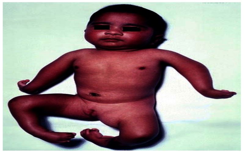
Which of the following is a characteristic feature of Arthrogryposis Multiplex Congenita (AMC)?
-
Limbs exhibit normal muscle tone and bulk.
-
Joints are contracted in flexion or extension.
-
The spine is typically straight without abnormal curvature.
-
Feet are commonly in the plantarflexion position.
Explanation
Correct Answer: B) Joints are contracted in flexion or extension.
Arthrogryposis Multiplex Congenita (AMC) is characterized by multiple joint contractures at birth. The affected joints can be contracted either in flexion or extension. Classic manifestations include flexed wrists, extended elbows, and knees, and hip dislocation with slight flexion. The limbs tend to be tubular and featureless, with hypoplastic (underdeveloped) muscles. This joint contracture is the hallmark feature of AMC.
Why Other Options are Wrong:
A) Limbs exhibit normal muscle tone and bulk.
This is incorrect. In AMC, muscle tone is typically reduced (hypotonia), and the muscles are hypoplastic (underdeveloped). The limbs appear featureless, and muscle bulk is often diminished, which is in contrast to the normal muscle tone and bulk suggested in this option.
C) The spine is typically straight without abnormal curvature.
This is incorrect. Scoliosis (abnormal curvature of the spine) can be present in AMC, not a straight spine. The condition often involves deformities not only in the joints but also in the spine.
D) Feet are commonly in the plantarflexion position.
This is incorrect. The feet in AMC are often in the equinovarus position (a type of clubfoot), not in plantarflexion. The equinovarus position is characterized by both inversion (turning inward) and plantarflexion (pointing downward) of the feet.
Summary:
Arthrogryposis Multiplex Congenita (AMC) is a congenital condition characterized by multiple joint contractures, typically in flexion or extension. It involves muscle hypoplasia and reduced tone. The condition can result in deformities like scoliosis and equinovarus feet. Early diagnosis and physical therapy are essential to manage joint mobility and muscle development.
In hypertensive emergencies, which organ systems are most commonly affected by marked blood pressure elevation?
-
Brain, Eyes, Heart, Kidneys
-
Brain, Lungs, Heart, Kidneys
-
Liver, Brain, Kidneys, Lungs
-
Heart, Kidneys, Lungs, Skin
Explanation
Correct Answer: A. Brain, Eyes, Heart, Kidneys
Marked blood pressure elevation in hypertensive emergencies can critically affect multiple organ systems. The most common and critical systems involved are:
Brain: Hypertensive encephalopathy, which can lead to seizures, coma, and altered mental status due to the damage from elevated pressure on cerebral blood vessels.
Eyes: Retinal changes, such as retinopathy, which can be seen in the retina during fundoscopic examination. This may include hemorrhages, exudates, and papilledema.
Heart: The heart can be impacted, with left ventricular heart failure being a common complication of uncontrolled hypertension, often due to the heart’s struggle to overcome the elevated pressure.
Kidneys: Renal insufficiency or acute kidney injury can result from sustained high blood pressure, as it leads to damage to the kidney vasculature and glomeruli. Elevated creatinine is typically a sign of renal damage.
Why the other options are wrong:
B. Brain, Lungs, Heart, Kidneys: While the brain, heart, and kidneys are correct, the lungs are not typically the primary system affected by hypertensive emergencies. The lungs are more involved in conditions like pulmonary edema related to heart failure but are not a primary concern in hypertensive emergencies.
C. Liver, Brain, Kidneys, Lungs: This option is incorrect because the liver is not one of the primary organs directly affected by hypertensive emergencies. Although high blood pressure can have secondary effects on the liver, it is not the most common or critical organ in hypertensive crises.
D. Heart, Kidneys, Lungs, Skin: This option is also incorrect as the skin is not typically affected by hypertensive emergencies. Skin issues may arise from secondary causes like medications or infections but are not a direct consequence of hypertension.
Summary:
In hypertensive emergencies, the most common and critical organ systems affected are the brain (hypertensive encephalopathy), eyes (retinal changes), heart (left ventricular heart failure), and kidneys (renal insufficiency). Marked blood pressure elevation can cause severe damage to these organs and often leads to life-threatening complications, making prompt recognition and treatment critical. Therefore, A is the correct answer.
How to Order
Select Your Exam
Click on your desired exam to open its dedicated page with resources like practice questions, flashcards, and study guides.Choose what to focus on, Your selected exam is saved for quick access Once you log in.
Subscribe
Hit the Subscribe button on the platform. With your subscription, you will enjoy unlimited access to all practice questions and resources for a full 1-month period. After the month has elapsed, you can choose to resubscribe to continue benefiting from our comprehensive exam preparation tools and resources.
Pay and unlock the practice Questions
Once your payment is processed, you’ll immediately unlock access to all practice questions tailored to your selected exam for 1 month .
Frequently Asked Question
Absolutely! fail is by choice.
We recommend bookmarking key sections or taking notes while online. This way, you’ll have the information handy even when you’re offline.
We regularly update our materials to ensure they align with the latest course content and exam patterns. Stay prepared with the most up-to-date resources!
You can reach out to us through our support system at any time for help with your studies. We're here to assist you!
Yes, each explanation is written in clear, simple language with examples to help clarify complex concepts and improve your understanding.
Absolutely! You have the flexibility to study anytime, anywhere, at your own pace without any deadlines or pressure.
Once you subscribe, you will gain instant access to all practice questions and explanations through our user-friendly online platform.
Yes! All questions are crafted based on the actual content areas covered in the NUR 335 course, ensuring that you are studying relevant and accurate material.